Digital Mole Mapping Article »
Moles & Early Diagnosis
Moles and their early diagnosis can be found in the National Cancer Institute (NCI) booklet (NIH Publication No. 99-3133), which will help you learn more about common moles and unusual ones called dysplastic nevi or atypical moles. This booklet shows what moles look like and explains how they may be related to melanoma, a type of skin cancer. It describes the signs of melanoma and explains how you can check your skin for moles that might be cancerous. It also explains why and how you can protect your skin.
Cancer research has led to real progress against cancer — better survival and an improved quality of life. Through research, our knowledge about moles and cancers of the skin keeps increasing. We are finding new ways to prevent, detect, and treat cancer. The Cancer Information Service and the other sources of NCI information listed under “National Cancer Institute Information Resources” can provide the latest, most accurate information about moles, dysplastic nevi, and cancer. Publications mentioned in this booklet and others are available from the Cancer Information Service at 1-800-4-CANCER. Many NCI publications are also available on the Internet at the Web sites listed in the “National Cancer Institute Information Resources” section at the end of this booklet.
Body Mole Mapping
Whole Body Photography (body mapping) is rapidly gaining popularity as a tool for managing patients at high risk for melanoma. Many of the leading cancer centers and over sixty percent of dermatology residency programs in the United States employ this technique to aid in the early detection of suspicious lesions.
Body Mapping Module
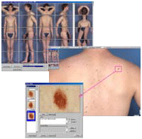
The Mirror Body Mapping module lets you link one or more close-up images to specific points on a body sector photo, making it easy to locate lesions on a patient’s body and track them over time. Ideal for pigmented lesions, body mapping is also useful for documenting psoriasis, eczema and other conditions.


